© Copyright 2020 Vanni VERONESI. All rights reserved.
Privacy Notice (ITALIANO) / Privacy Policy (ENGLISH)Cookie Notice (ITALIANO) / Cookies information (ENGLISH)
LUMBAR MAGNETIC RESONANCE IMAGING (MRI) IN PRONE POSITION IN
OCCULT TETHERED CORD SYNDROME
Association of lumbar MRI in prone position and minimally invasive surgery of FILUM TERMINALE EXTERNUM (EXTRADURAL) SECTION under local anaesthesia in OCCULT TETHERED CORD SYNDROME
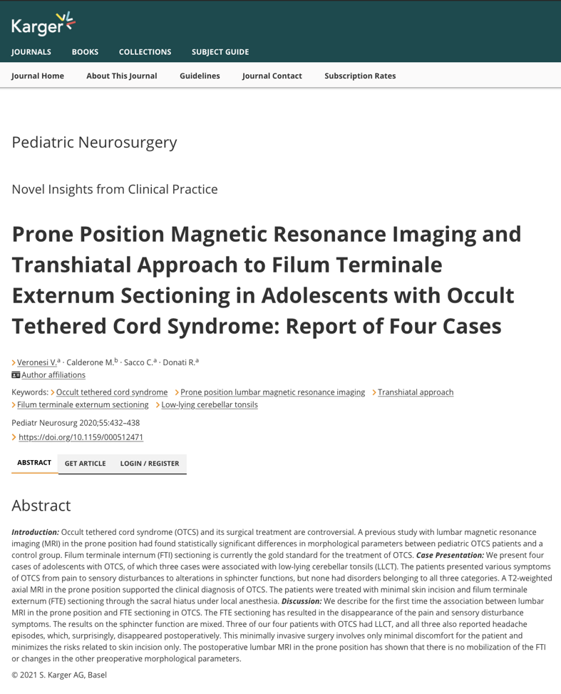
This scientific work of ours was published in the scientific journal Pediatric Neurosurgery in 2020 and concerns the association between prone lumbar MRI and extradural (externum) section of the filum terminale, under local anaesthesia, for the treatment of OCCULT TETHERED CORD syndrome.
We first described this association between prone lumbar MRI and minimally invasive surgery for the extradural section of the filum terminale in the international medical literature.
Patients operated on with this MINIMALLY INVASIVE SURGERY are generally adults and, to date, have numbered over 400; we selected four adolescent patients who were operated on, and these are the subjects of this scientific work.
We do not treat children.
LUMBAR MRI IN PRONE POSITION IN OCCULT TETHERED CORD and traditional surgery of INTRADURAL SECTION of the filum terminale
terminal under GENERAL ANESTHESIA
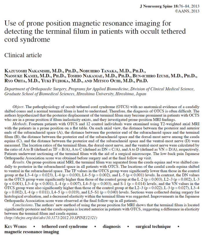
This 2013 scientific work was published in an international medical journal by Japanese colleagues at Hiroshima University.
MRI in the prone position was used to identify the abnormal position of the intradural filum terminale and nerve roots in patients with occult tethered cord syndrome.
The Japanese colleagues compared a group of patients with occult tethered cord syndrome with a group of asymptomatic individuals.
MRI in the prone position revealed a significant difference in the position of the filum terminale and nerve roots between the two groups.
In patients with occult tethered cord syndrome, the filum terminale did not gravitationally move anteriorly with the nerve roots, but remained posterior, and the nerve roots were positioned in the anterior part of the dural sac.
Obviously, determining the posterior position of the filum terminale does not allow us to assess the intrinsic elasticity of the filum terminale itself and, therefore, determine whether there are correlations with symptoms. However, a taut filum terminale can strain the spinal cord to which it is attached, causing the symptoms seen in OCCULT TETHERED CORD syndrome.
This pioneering scientific work, however, highlights a correlation between certain morphological and clinical findings.
Further studies will be needed to understand the specificity (ability to identify true negatives) and sensitivity (ability to identify true positives) of this particular MRI.
LUMBAR MRI IN PRONE POSITION IN THE TETHERED CORD
AND IN THE RETETHERING
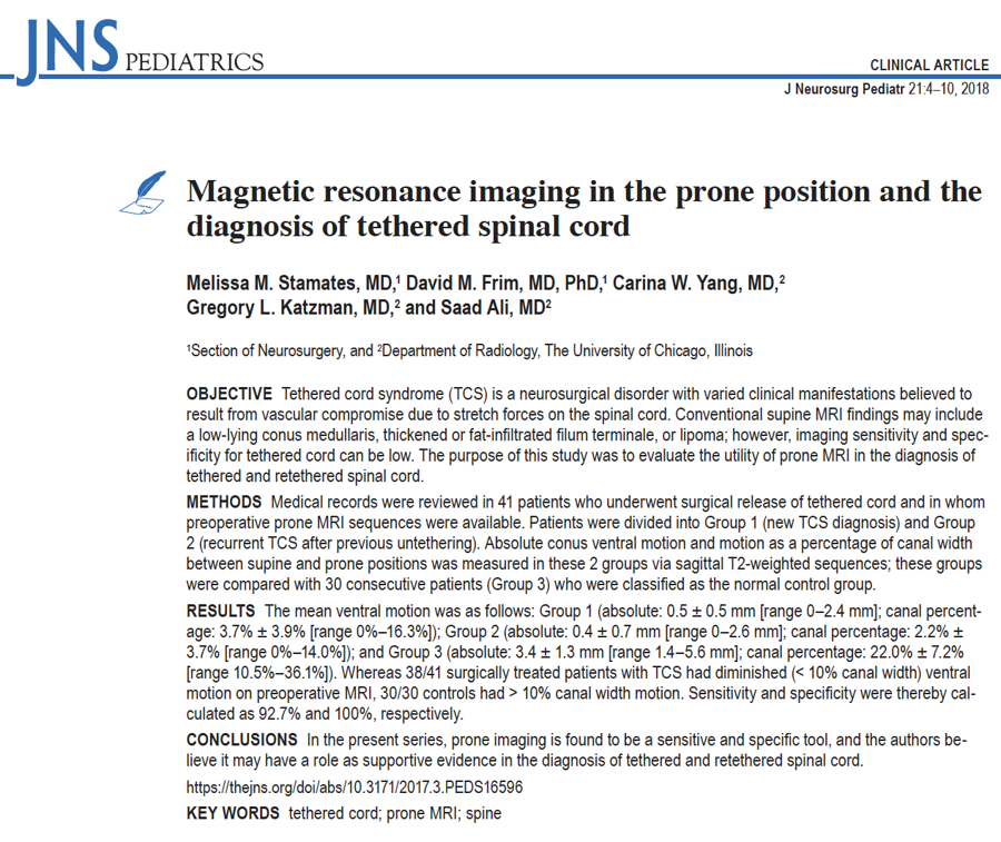
In 2018, American colleagues at the University of Chicago conducted a study to evaluate the usefulness of prone lumbar MRI in cases of tethered cord and spinal cord re-tethering.
They also evaluated a small group of patients with occult tethered cord, which, however, was not included in the case study analysis.
The American colleagues, evaluating the percentage of motion of the conus medullaris in the supine and prone positions, concluded their study by stating:
"In the present series, prone MRI proved to be a sensitive and specific tool, and the authors believe it may have a role as a supportive test in the diagnosis of tethered cord or spinal cord re-tethering."