© Copyright 2020 Vanni VERONESI. All rights reserved.
Privacy Notice (ITALIANO) / Privacy Policy (ENGLISH)Cookie Notice (ITALIANO) / Cookies information (ENGLISH)
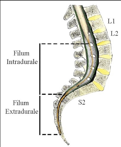
MINIMALLY INVASIVE SURGERY UNDER LOCAL ANESTHESIA FOR THE SECTION OF THE EXTRADURAL (EXTERNUM) FILUM TERMINALE
Minimally invasive surgery under local anaesthesia for sectioning the extradural filum terminale may present the only complication: a skin infection at the site of the surgical incision. To prevent this, the protocol calls for antibiotic prophylaxis in the operating room before the procedure.
The skin incision measures approximately 4 cm and is made at the level of the sacral hiatus. During this procedure, no bone is removed from the vertebrae or sacrum. Using microsurgical techniques under an operating microscope, the posterior sacrococcygeal ligament is removed, and the extradural filum terminale is isolated and cut. For a video description of the technique, CLICK HERE.
Because there are no meninges or nervous structures in the surgical field, there is no neurological damage or meningitis. The surgery lasts approximately 25 minutes, and the patient can resume walking approximately 60 minutes later. The patient is discharged from the hospital on the same day or the following day.
For anatomical reasons, the procedure does not involve opening the meninges. Therefore, this minimally invasive surgical technique, in addition to avoiding the complications of traditional surgery involving intradural division of the filum terminale (for a list, see the next post), eliminates the risk of re-anchoring associated with opening the meninges, which is typical of traditional surgery under general anaesthesia.
ADVANTAGES of minimally invasive surgery for the EXTRADURAL section of the FILUM TERMINALE under local anaesthesia
- LOCAL ANESTHESIA
- SHORT PROCEDURE, APPROXIMATELY 25 MINUTES.
- SMALL SKIN CUT, APPROXIMATELY 4 CENTIMETERS (see box below); smaller wounds expose the body less to potential hospital-acquired pathogens.
- ELECTROPHYSIOLOGICAL MONITORING IS NOT NECESSARY DURING SURGERY, SINCE THERE ARE NO NERVE STRUCTURES IN THE OPERATING FIELD; THEREFORE, THERE IS NO RISK OF NEUROLOGICAL COMPLICATIONS.
- BY NOT OPENING THE MENINGES, MENINGITIS IS NOT A POSSIBLE COMPLICATION.
- AFTER SURGERY, ONLY ONE HOUR OF BED REST IS REQUIRED.
- HOSPITAL DISCHARGE POSSIBLE ON THE DAY OF SURGERY.
- THE ANATOMICAL CONDITIONS FOR RE-ANCHORAGE ARE NOT PRESENT.
A small skin incision is sufficient to perform the minimally invasive surgical approach to the sacral spinal canal through the SACRAL HIATUS for the section of the EXTRADURAL FILUM TERMINALE
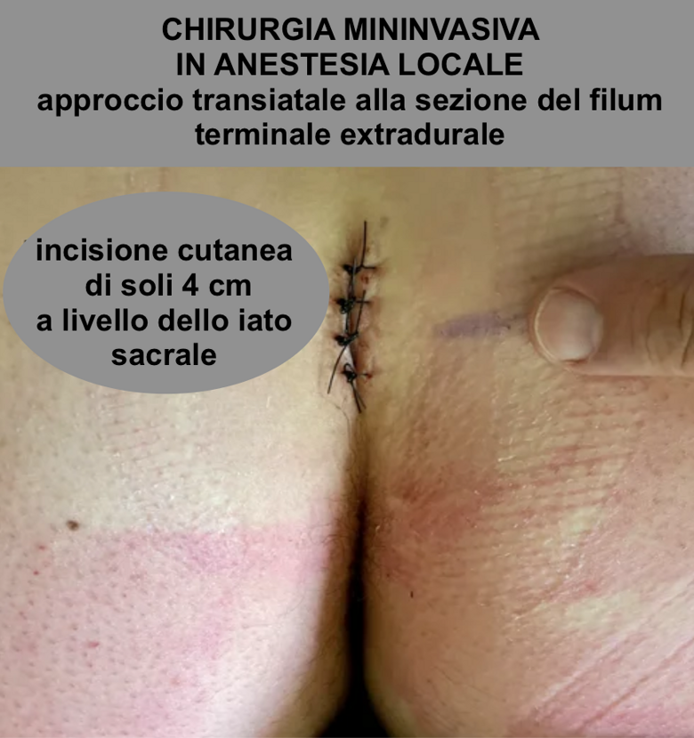
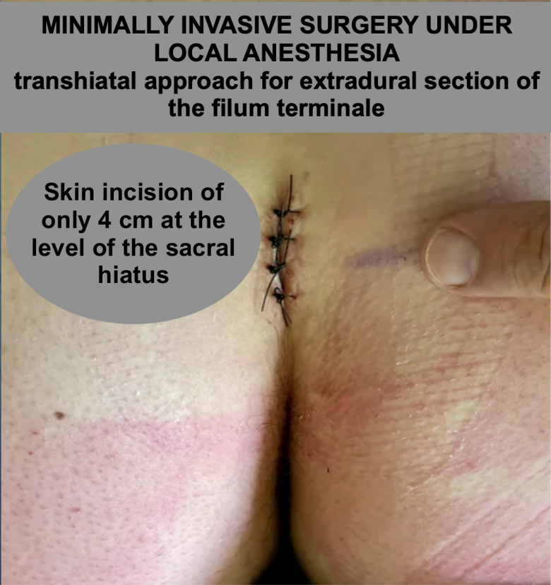
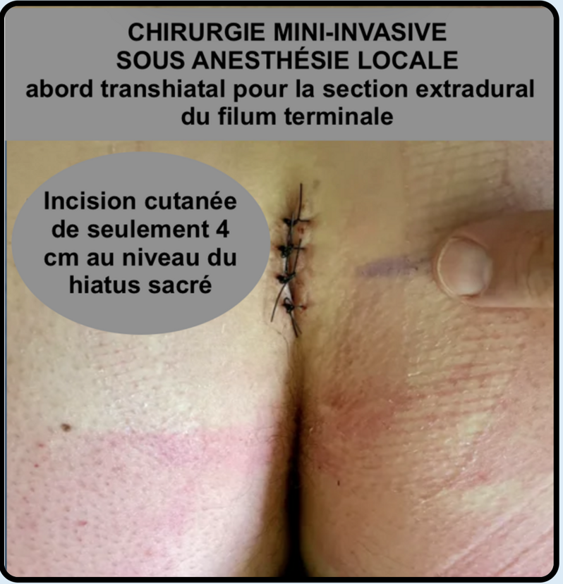

Landmark for transhiatal approach to the EXTRADURAL filum terminale.
Two posterior superior iliac crests form an equilateral triangle.
The top of this triangle indicates the V-shaped SACRAL HIATUS, limited by two sacral cornua.
INFORMATIONAL VIDEO
SECTIONING OF EXTRADURAL FILUM TERMINALE UNDER LOCAL ANESTHESIA:
- ANATOMY
- SURGICAL TECHNIQUE
- POSTOPERATIVE COURSE
FOR SCIENTIFIC AND INFORMATIONAL PURPOSES, CLICK BELOW TO CONNECT TO THE YOUTUBE CHANNEL OF VERONESI VANNI AND WATCH THE VIDEO
SURGICAL VIDEO

SECTIONING OF EXTRADURAL (EXTERNUM) FILUM TERMINALE UNDER LOCAL ANESTHESIA
In the video, the Brazilian patient is awake and speaks with the interpreter Prof. Giorgio Charissiadis, who describes the surgical steps to her.
The operating microscope is used in the central part of the surgical procedure for the sectioning of the extradural filum terminale.
TRADITIONAL SURGERY UNDER GENERAL ANESTHESIA FOR THE SECTIONING OF THE INTRADURAL FILUM TERMINALE
Traditional intradural sectioning of the filum terminale requires general anaesthesia for 1-2 hours.
The complication rate is very low, but the risks of traditional intradural sectioning of the filum terminale under general anaesthesia include possible permanent urinary and/or faecal sphincter control deficits and new neurological deficits.
There is a risk of cerebrospinal fluid leakage with the development of a fistula or pseudomeningocele, or tethering of the cauda equina and conus medullaris, which may require subsequent surgery.
There is a risk of wound healing problems and meningitis, with associated morbidity and mortality.
The traditional surgical approach involves:
- removal of the posterior aspect of the vertebrae;
- opening of the meninges;
- identification of the intradural filum terminale between the nerve roots of the cauda equina (and verification with electrical stimulation to avoid severing the nerve roots and resulting neurological damage).
The procedure lasts approximately 60-90 minutes.
Before starting the procedure, which is always under general anaesthesia, the necessary time is added to set up intraoperative electrophysiological monitoring.
The length of the skin incision is usually between 8 and 12 cm.
After surgery, bed rest in the prone position is recommended for 1-2 days.
Discharge is expected within three to four days of surgery.
Cases of re-anchoring due to adhesions of the previously opened meninges have been described with this technique.